
World-Class Medical Technology,
Powered by More Precise Radiotherapy Science.
Modern precision radiotherapy is not simply about “directing radiation at a tumor.” From target identification and dose design to normal-tissue protection, respiratory motion management and anatomical changes during treatment, every step can influence the final treatment plan. Even patients with the same cancer diagnosis may require very different radiotherapy techniques and treatment platforms.
The Goal of Precision Radiotherapy Is Not Simply to Deliver More Radiation into the Body. Its real technical value lies in shaping the treatment dose around the tumor’s three-dimensional anatomy and biological needs, while limiting radiation exposure to surrounding normal tissues as much as possible. Next, we will begin with the principles of radiotherapy and explore why Photon, Proton and different treatment platforms exist.
How Does Radiotherapy
Actually Affect Cancer Cells?
Radiotherapy does not simply “burn away” a tumor. High-energy radiation or particles deposit energy in tissue, causing DNA damage within cells. If cancer cells cannot repair this damage effectively, they gradually lose the ability to continue dividing.
From Radiation Entering the Body to Cancer Cells Losing the Ability to Proliferate.
Modern radiotherapy focuses on where energy is deposited, how much dose the tumor receives and how much unnecessary dose is delivered to normal tissues.
More Radiotherapy Sessions Are Not Necessarily Better, and Fewer Sessions Are Not Necessarily More Advanced.
Dose per fraction, total dose, cancer type, biological characteristics, lesion location and the tolerance of surrounding normal tissues all influence the choice of fractionation schedule.
Cancer type, growth rate and radiosensitivity can influence dose and fractionation design.
Treatment planning must also consider dose limits for normal organs such as the spinal cord, brainstem, lungs and bowel.
The dose prescription comes from clinical judgment, then the appropriate treatment platform is selected to deliver the plan.
The Most Important Difference with Proton Therapy
Is Not That It Is “Stronger,” but Where the Dose Stops.
Both photons and protons can be used to treat tumors, but they deposit energy inside the body in different ways. A key physical characteristic of proton therapy is the Bragg Peak, which allows a higher dose to be concentrated near a selected depth while the distal dose falls rapidly.
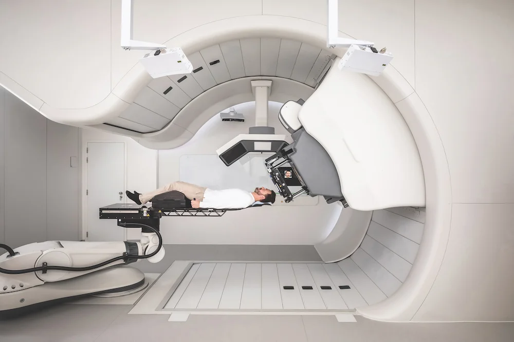
The Biggest Difference Between Photon and Proton Therapy Often Appears “Beyond the Tumor.”
Both can cover the tumor with treatment dose, but what happens to the dose after it leaves the target is an important distinction.
Photon Radiotherapy
After X-rays enter the body, they continue depositing energy along their path; even after passing through the tumor, a certain amount of exit dose remains.
Proton Therapy
By controlling proton energy, the higher dose can be concentrated near a planned depth and fall rapidly beyond the target.
For pediatric tumors, lesions near the skull base or central nervous system, and selected tumors close to critical organs, doctors may pay particular attention to whether a proton plan can reduce dose to surrounding normal tissues.
What should actually be compared is tumor coverage, dose to normal organs, lesion motion, treatment robustness and the overall clinical objective, rather than simply comparing the names of treatment machines.
Why Does One Hospital Need
So Many Different Radiotherapy Systems?
Because “radiotherapy” is not a single fixed technology. Small lesions, moving targets, complex extended treatment areas, and tumors affected by daily anatomical changes present very different technical challenges. The real value lies in having multiple platforms to match different clinical needs.
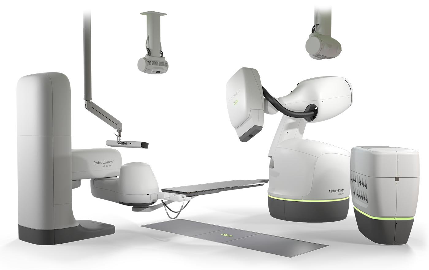
CyberKnife M6
A robotic arm delivers highly precise radiation beams from multiple angles, combined with imaging and tracking technologies for small, complex or moving lesions.
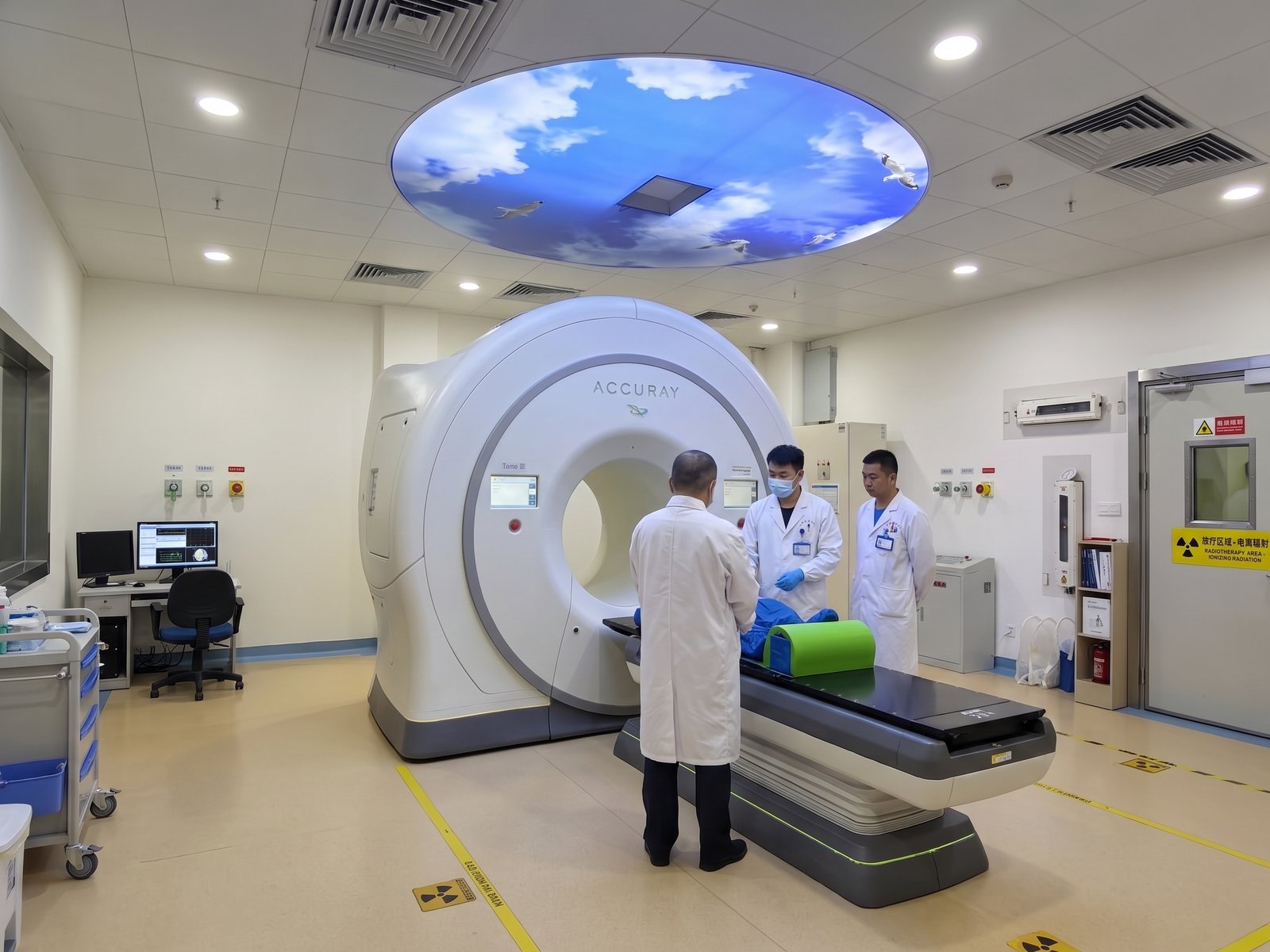
TomoTherapy
The gantry rotates while the treatment couch moves, delivering radiation in a helical pattern for complex shapes, extended treatment fields or multiple targets.
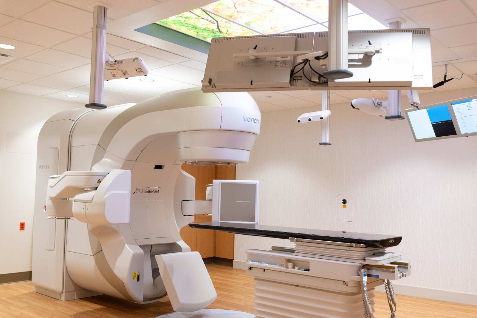
TrueBeam
A versatile platform supporting IMRT, VMAT, IGRT, SRS and SBRT for a wide range of precision radiotherapy needs.
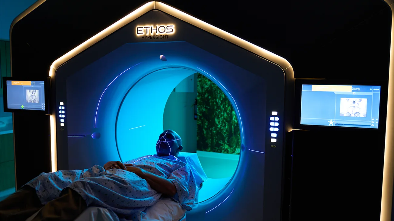
Ethos
When organ positions or tumor anatomy change from day to day, online adaptive workflows can be performed using the day’s imaging.
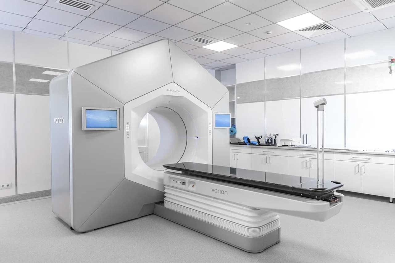
Halcyon + HyperSight
A high-efficiency image-guided platform combined with faster, higher-quality HyperSight treatment imaging.
Not All Radiotherapy Comes from an External Linear Accelerator.
Some tumors may also be treated with brachytherapy, intraoperative radiotherapy or other specialized techniques, bringing the radiation source closer to the treatment area or delivering a local dose directly during surgery.
A Machine Name Is Not the Same as an Indication.
The same machine can deliver multiple radiotherapy techniques, while the same cancer type may require a completely different platform depending on the lesion characteristics.
What We Treat
Is Not a Static CT Image.
Breathing can move lung and liver lesions, bladder and rectal filling can alter pelvic anatomy, and the tumor itself may shrink over several weeks of treatment. Modern radiotherapy therefore requires more than accurate positioning on the first day; the patient’s actual anatomy must continue to be verified throughout treatment.
When the Body Changes Day by Day, the Treatment Plan Can Change with It.
The core of online adaptive radiotherapy is not to “redo radiotherapy every day,” but to use the day’s imaging to reassess the target and normal organs, and re-optimize the plan when needed.
Lung and Liver Lesions Can Move with Breathing.
If a treatment plan is based on only one moment in time, the true range of motion may be underestimated. Selected cases may therefore require motion-management approaches such as 4D-CT, respiratory gating or dynamic tracking.
The Patient’s Anatomy Is Not Exactly the Same Every Day.
Bladder filling, rectal gas, weight changes and shifts in organ position can all affect the spatial relationship between the target and surrounding normal tissues.
The Tumor May Also Shrink During Several Weeks of Treatment.
When tumor volume or the relationship between organs changes significantly, the original plan may no longer be the most appropriate. Adaptive techniques provide an opportunity to reassess and optimize the plan.
The exact clinical workflow varies by equipment, disease type and treatment plan; the steps below illustrate the core logic.
Faster Imaging Is Not Only About Saving Time.
Faster, higher-quality treatment imaging can help clarify the relationship between the target and normal organs on that day, while providing a stronger imaging basis for adaptive decisions.
Multiple Brain Metastases Do Not Necessarily Need to Be Treated One by One.
Using a single isocenter and non-coplanar arc delivery, HyperArc can treat multiple intracranial targets within one treatment plan while balancing treatment efficiency and dose distribution.
True Technical Capability
Is Built Through Clinical Experience Accumulated Every Day.
Radiotherapy equipment is only a tool. What truly determines treatment quality is whether imaging, target contouring, dose design, treatment verification, motion management and clinical judgment can be executed consistently in everyday practice.

From Introducing New Technology to Building Consistent Clinical Experience.
Advanced technology only gains practical meaning when it becomes part of routine clinical workflows and is supported by continuous case accumulation, quality control and team experience.
Already Have a Radiotherapy Recommendation but Unsure Which Technique Is More Suitable?
You can submit your current pathology, recent CT / MRI / PET imaging and previous radiotherapy records so the medical team can first assess the treatment objective, target characteristics and normal-tissue risks before evaluating the most appropriate radiotherapy approach.
True Precision
Is Not About Choosing the Most Expensive or Newest Machine.
It means understanding the cancer type, imaging, target, previous treatment, normal-organ tolerance and treatment objective first — then selecting the most appropriate dose, fractionation schedule and technology platform for this specific course of treatment.
Can Radiotherapy Be Given Again After a Previous Course?
Re-irradiation may be considered for selected patients, but the decision cannot be based simply on whether radiotherapy was given before. Doctors need to reassess the previous dose and treatment field and estimate the cumulative dose already received by critical normal tissues.
Understand the Tumor First, Then Choose the Technology.
Even with the same cancer diagnosis, patients may differ in lesion location, size, number, motion, previous radiotherapy history and treatment goals. Therefore, there is no single fixed answer that makes Proton, CyberKnife, TomoTherapy, TrueBeam, Halcyon or Ethos the best choice for everyone.
The Most Advanced Technology Is Not Necessarily What Every Patient Needs;
The Most Appropriate Plan Is the Real Goal of Precision Radiotherapy.
The machine is selected after the clinical problem is understood — not the other way around.
About Radiotherapy,
These Are the Questions Patients Ask Most Often.
The following information is intended to help patients understand common radiotherapy concepts. The actual treatment dose, number of sessions and technology selection still require individualized assessment based on pathology, imaging, previous treatment and risks to normal organs.
01Will Radiotherapy Also Damage Normal Cells?
02Why Do Some Patients Need Only 5 Sessions While I Need 25–30?
03Which Is Best: CyberKnife, TomoTherapy or TrueBeam?
04Is Proton Therapy Always Better Than Conventional Radiotherapy?
05What Happens If the Tumor Shrinks During Radiotherapy?
06If Lung or Liver Tumors Move with Breathing, How Is Radiotherapy Kept Accurate?
07If I Have Already Had Radiotherapy, Can I Receive It Again?
08If I Am Overseas, Can the Team First Assess Which Radiotherapy May Be Suitable?
Want to Know Which Radiotherapy Technique May Be More Suitable for Your Case?
You can submit your pathology, recent imaging and previous treatment records so the medical team can make an initial assessment based on treatment goals, target characteristics and risks to normal tissues.